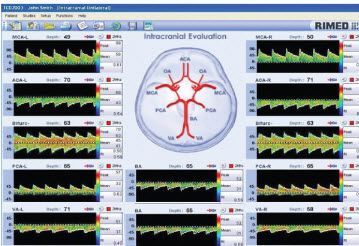

As fluid flows from a conduit (or blood vessel) of greater diameter to one of smaller diameter, the flow velocity must increase to allow the same volume of blood to flow through the narrower area (this is analogous to one putting one’s thumb over the opening of a garden hose as water is flowing out resulting in a high-velocity jet of water being formed). According to the principle, as fluid flows through conduits of differing diameters, the velocity and pressure associated with the fluid will change. At our institution, spectral Doppler waveforms and flow velocities are obtained from the proximal, mid, and distal MCA and single measurements are obtained in the visualized portion of the ACA, PCA, and terminal ICA.īernoulli’s principle, as applied to flowing liquids, can be utilized for the diagnosis of areas of stenosis and vasospasm. With the transtemporal approach, visualization of the MCA, ACA, PCA, and terminal ICA is possible. Flow velocity measurements (both MFV and peak systolic velocity) are obtained. Imaging is performed from the left and right temporal bones with color Doppler flow imaging utilized to visualize the vessels.

The transducer is placed on the temporal bone either above the zygomatic arch and anterior to the external auditory canal or slightly more cephalad and posterior over the earlobe. Imaging of the circle of Willis is performed via a transtemporal approach utilizing a 5–1 MHz sector-array transducer with most of the imaging being performed at the lower frequencies (1–2 MHz) to allow penetration of the temporal bone. With advances in ultrasound equipment and the utilization of gray-scale, color Doppler flow, and spectral Doppler imaging, direct visualization of the vessel and more accurate flow velocity measurements are now possible.


 0 kommentar(er)
